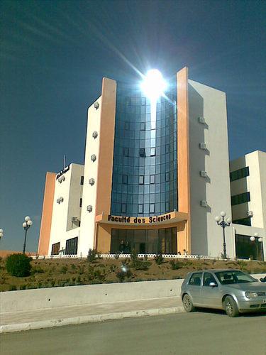
University SÃĐtif 1 FERHAT ABBAS Faculty of Sciences
DÃĐtail de l'auteur
Auteur Adouda Adjiri |
Documents disponibles écrits par cet auteur


 Ajouter le rÃĐsultat dans votre panier Affiner la recherche
Comparaison entre les sÃĐquences d'IRM pour la dÃĐtection des lÃĐsions hÃĐpatiques focales / Yousra Benimeur

Titre : Comparaison entre les sÃĐquences d'IRM pour la dÃĐtection des lÃĐsions hÃĐpatiques focales Type de document : texte imprimÃĐ Auteurs : Yousra Benimeur, Auteur ; Adouda Adjiri, Directeur de thÃĻse AnnÃĐe de publication : 2023 Importance : 1 vol (51 f.) Format : 29 cm Langues : Français (fre) CatÃĐgories : Physique Mots-clÃĐs : Physique Index. dÃĐcimale : 530-Physique Côte titre : MAPH/0598 En ligne : https://drive.google.com/file/d/1nEFseOoTldU1XWtV_krNCu2Ph57L7LzP/view?usp=shari [...] Format de la ressource ÃĐlectronique : Comparaison entre les sÃĐquences d'IRM pour la dÃĐtection des lÃĐsions hÃĐpatiques focales [texte imprimÃĐ] / Yousra Benimeur, Auteur ; Adouda Adjiri, Directeur de thÃĻse . - 2023 . - 1 vol (51 f.) ; 29 cm.
Langues : Français (fre)
CatÃĐgories : Physique Mots-clÃĐs : Physique Index. dÃĐcimale : 530-Physique Côte titre : MAPH/0598 En ligne : https://drive.google.com/file/d/1nEFseOoTldU1XWtV_krNCu2Ph57L7LzP/view?usp=shari [...] Format de la ressource ÃĐlectronique : Exemplaires (1)
Code-barres Cote Support Localisation Section DisponibilitÃĐ MAPH/0598 MAPH/0598 Mémoire Bibliothèque des sciences Français Disponible
DisponibleEvaluation of ADC ratio in Diffusion-Weighted Imaging: Application for liver focal lesions and brain tumors / Houda Zine
Titre : Evaluation of ADC ratio in Diffusion-Weighted Imaging: Application for liver focal lesions and brain tumors Type de document : document ÃĐlectronique Auteurs : Houda Zine, Auteur ; Meriem Mekideche, Auteur ; Adouda Adjiri, Directeur de thÃĻse Editeur : Setif:UFA AnnÃĐe de publication : 2024 Importance : 1 vol (37 f.) Format : 29 cm Langues : Anglais (eng) CatÃĐgories : ThÃĻses & MÃĐmoires:Physique Mots-clÃĐs : Diffusion-weighted imaging (DWI)
Apparent diffusion coefficient (ADC)
Liver lesions
Brain tumors
B value
Region of interest (ROI).Index. dÃĐcimale : 530 - Physique RÃĐsumÃĐ :
Diffusion-weighted imaging or DWI is based on the study of the diffusion of water molecules between and within cells. This technique provides information about the cellular structure of tissues by calculating the apparent diffusion coefficient or ADC; thereby quantifying the rate of water diffusion and providing information on tissue microstructure and cellularity. The objective is to generate ADC maps with reliable values aiming to differentiate between benign and malignant lesions.
We studied a total of 31 patients with liver lesions or brain tumors. As part of this study we noted that it is possible to differentiate a benign lesion from a malignant lesion using the ADC value but the age of the patient must be taken into consideration because it plays a major role. We also noted that there are several other factors which affect the value of ADC such as the image acquisition parameters, including the choice of the (b) value and the region of interest (ROI). This affirms the important role that a medical physicist must play in mastering the physical parameters linked to the generation of DW images that are of clinical value and useful to calculate reliable ADC values.Note de contenu :
Sommaire
Chapitre I. Diffusion-Weighted Imaging ..................................................................................... 2
I.1. Introduction ...................................................................................................................... 2
I.2. Diffusion .......................................................................................................................... 2
I.2.1. Isotropic diffusion ...................................................................................................... 2
I.2.2. Anisotropic diffusion ................................................................................................. 3
I.2.3. Restricted diffusion .................................................................................................... 3
I.3. Diffusion tensor ................................................................................................................ 3
I.4. Diffusion -Weighted Imaging ........................................................................................... 4
I.4.1. Definition .................................................................................................................. 4
I.4.2. The b value ................................................................................................................ 5
I.4.3. Apparent Diffusion Coefficient .................................................................................. 7
Chapitre II. Focal liver and Brain lesions .................................................................................... 8
II.1. Introduction ..................................................................................................................... 8
II.2. Anatomy ......................................................................................................................... 8
II.2.1. Liver Anatomy ......................................................................................................... 8
II.2.2. Brain Anatomy ......................................................................................................... 8
II.3. Liver and brain lesions .................................................................................................... 9
II.3.1. Focal liver lesions ..................................................................................................... 9
II.3.2. Brain lesions ........................................................................................................... 10
II.4. Hepatic and brain MRI Sequences ................................................................................. 10
II.5. Image quality criteria..................................................................................................... 10
II.5.1. The signal-to-noise ratio ......................................................................................... 11
II.5.2. Contrast: ................................................................................................................. 12
II.5.3. Spatial resolution: ................................................................................................... 12
II.5.4. Acquisition time: .................................................................................................... 12
II.6. Conclusion: ................................................................................................................... 12
Chapitre III. Experimental part ................................................................................................. 13
III.1. Introduction ................................................................................................................. 13
III.2. When is the diagnosis made using MRI ........................................................................ 13
III.3. Methods ....................................................................................................................... 13
III.3.1. Imaging technique ................................................................................................. 13
III.3.2. Example of DWI, b0 and image of a human brain ................................................. 14
III.4. Results ......................................................................................................................... 14
III.4.1. Parameters that affect DWI image quality ............................................................. 14
III.4.1.1. The effect of spatial resolution ........................................................................ 14
III.4.1.1.1 The effect of the matrix size on spatial resolution ................................... 14
III.4.1.1.2 The effect of slice thickness and gap ...................................................... 16
III.4.1.2. The effect of b Value on image quality ........................................................... 16
III.4.1.2.1 Case of the brain .................................................................................... 16
III.4.1.2.2 Case of the liver ..................................................................................... 16
III.4.1.3. The effect of excitation number on image quality ........................................... 18
III.4.1.4. The effect of TE and TR on image quality ...................................................... 18
III.4.2. Parameters that affect ADC value and image quality ............................................. 19
III.4.2.1. Effect of the choice of the region of interest (ROI).......................................... 19
III.4.2.2. The effect of b value on the ADC value .......................................................... 20
III.4.2.3. The effect of ROI effect on the ADC value in normal and tumor tissue ........... 21
III.4.2.4. The effect of spatial resolution on ADC value................................................. 22
III.4.2.4.1 The effect of the matrix on ADC value ................................................... 22
III.4.2.4.2 The effect of thickness on ADC value and image quality ........................ 22
III.4.2.5. The effect of Nex on ADC value .................................................................... 23
III.4.2.6. The effect of TE and TR on ADC value .......................................................... 24
III.4.3. ADC Ratio to differentiate between benign and malignant lesions in the liver and brain ................................................................................................................................ 24
III.4.3.1. Steps to take the ADC value and determine the ROI ....................................... 24
III.4.3.1.1 . Ensuring the homogeneity of the lesion ................................................ 24
III.4.3.1.2 Determining the center of the region of interest ...................................... 24
III.4.3.2. ADC Ratio in benign and malignant tissues .................................................... 26
A. Liver ............................................................................................................................... 26
B. Brain ............................................................................................................................... 28Côte titre : MAPH/0661 Evaluation of ADC ratio in Diffusion-Weighted Imaging: Application for liver focal lesions and brain tumors [document ÃĐlectronique] / Houda Zine, Auteur ; Meriem Mekideche, Auteur ; Adouda Adjiri, Directeur de thÃĻse . - [S.l.]Â : Setif:UFA, 2024 . - 1 vol (37 f.) ; 29 cm.
Langues : Anglais (eng)
CatÃĐgories : ThÃĻses & MÃĐmoires:Physique Mots-clÃĐs : Diffusion-weighted imaging (DWI)
Apparent diffusion coefficient (ADC)
Liver lesions
Brain tumors
B value
Region of interest (ROI).Index. dÃĐcimale : 530 - Physique RÃĐsumÃĐ :
Diffusion-weighted imaging or DWI is based on the study of the diffusion of water molecules between and within cells. This technique provides information about the cellular structure of tissues by calculating the apparent diffusion coefficient or ADC; thereby quantifying the rate of water diffusion and providing information on tissue microstructure and cellularity. The objective is to generate ADC maps with reliable values aiming to differentiate between benign and malignant lesions.
We studied a total of 31 patients with liver lesions or brain tumors. As part of this study we noted that it is possible to differentiate a benign lesion from a malignant lesion using the ADC value but the age of the patient must be taken into consideration because it plays a major role. We also noted that there are several other factors which affect the value of ADC such as the image acquisition parameters, including the choice of the (b) value and the region of interest (ROI). This affirms the important role that a medical physicist must play in mastering the physical parameters linked to the generation of DW images that are of clinical value and useful to calculate reliable ADC values.Note de contenu :
Sommaire
Chapitre I. Diffusion-Weighted Imaging ..................................................................................... 2
I.1. Introduction ...................................................................................................................... 2
I.2. Diffusion .......................................................................................................................... 2
I.2.1. Isotropic diffusion ...................................................................................................... 2
I.2.2. Anisotropic diffusion ................................................................................................. 3
I.2.3. Restricted diffusion .................................................................................................... 3
I.3. Diffusion tensor ................................................................................................................ 3
I.4. Diffusion -Weighted Imaging ........................................................................................... 4
I.4.1. Definition .................................................................................................................. 4
I.4.2. The b value ................................................................................................................ 5
I.4.3. Apparent Diffusion Coefficient .................................................................................. 7
Chapitre II. Focal liver and Brain lesions .................................................................................... 8
II.1. Introduction ..................................................................................................................... 8
II.2. Anatomy ......................................................................................................................... 8
II.2.1. Liver Anatomy ......................................................................................................... 8
II.2.2. Brain Anatomy ......................................................................................................... 8
II.3. Liver and brain lesions .................................................................................................... 9
II.3.1. Focal liver lesions ..................................................................................................... 9
II.3.2. Brain lesions ........................................................................................................... 10
II.4. Hepatic and brain MRI Sequences ................................................................................. 10
II.5. Image quality criteria..................................................................................................... 10
II.5.1. The signal-to-noise ratio ......................................................................................... 11
II.5.2. Contrast: ................................................................................................................. 12
II.5.3. Spatial resolution: ................................................................................................... 12
II.5.4. Acquisition time: .................................................................................................... 12
II.6. Conclusion: ................................................................................................................... 12
Chapitre III. Experimental part ................................................................................................. 13
III.1. Introduction ................................................................................................................. 13
III.2. When is the diagnosis made using MRI ........................................................................ 13
III.3. Methods ....................................................................................................................... 13
III.3.1. Imaging technique ................................................................................................. 13
III.3.2. Example of DWI, b0 and image of a human brain ................................................. 14
III.4. Results ......................................................................................................................... 14
III.4.1. Parameters that affect DWI image quality ............................................................. 14
III.4.1.1. The effect of spatial resolution ........................................................................ 14
III.4.1.1.1 The effect of the matrix size on spatial resolution ................................... 14
III.4.1.1.2 The effect of slice thickness and gap ...................................................... 16
III.4.1.2. The effect of b Value on image quality ........................................................... 16
III.4.1.2.1 Case of the brain .................................................................................... 16
III.4.1.2.2 Case of the liver ..................................................................................... 16
III.4.1.3. The effect of excitation number on image quality ........................................... 18
III.4.1.4. The effect of TE and TR on image quality ...................................................... 18
III.4.2. Parameters that affect ADC value and image quality ............................................. 19
III.4.2.1. Effect of the choice of the region of interest (ROI).......................................... 19
III.4.2.2. The effect of b value on the ADC value .......................................................... 20
III.4.2.3. The effect of ROI effect on the ADC value in normal and tumor tissue ........... 21
III.4.2.4. The effect of spatial resolution on ADC value................................................. 22
III.4.2.4.1 The effect of the matrix on ADC value ................................................... 22
III.4.2.4.2 The effect of thickness on ADC value and image quality ........................ 22
III.4.2.5. The effect of Nex on ADC value .................................................................... 23
III.4.2.6. The effect of TE and TR on ADC value .......................................................... 24
III.4.3. ADC Ratio to differentiate between benign and malignant lesions in the liver and brain ................................................................................................................................ 24
III.4.3.1. Steps to take the ADC value and determine the ROI ....................................... 24
III.4.3.1.1 . Ensuring the homogeneity of the lesion ................................................ 24
III.4.3.1.2 Determining the center of the region of interest ...................................... 24
III.4.3.2. ADC Ratio in benign and malignant tissues .................................................... 26
A. Liver ............................................................................................................................... 26
B. Brain ............................................................................................................................... 28Côte titre : MAPH/0661 Exemplaires (1)
Code-barres Cote Support Localisation Section DisponibilitÃĐ MAPH/0661 MAPH/0661 Mémoire Bibliothèque des sciences Anglais Disponible
Disponible

