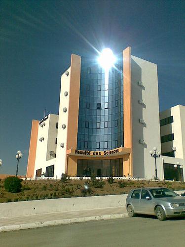
University SÃĐtif 1 FERHAT ABBAS Faculty of Sciences
DÃĐtail de l'auteur
Auteur Naoures Nourhane Khellaf |
Documents disponibles écrits par cet auteur


 Ajouter le rÃĐsultat dans votre panier Affiner la recherche
Design and 3D printing of dedicated phantom for the collection of radiotherapy data on PLA dose bolus material / Naoures Nourhane Khellaf
Titre : Design and 3D printing of dedicated phantom for the collection of radiotherapy data on PLA dose bolus material Type de document : document ÃĐlectronique Auteurs : Naoures Nourhane Khellaf, Auteur ; Fayçal Kharfi, Directeur de thÃĻse Editeur : Setif:UFA AnnÃĐe de publication : 2024 Importance : 1 vol (64 f.) Format : 29 cm Langues : Français (fre) CatÃĐgories : ThÃĻses & MÃĐmoires:Physique Mots-clÃĐs : Radiotherapy Reference data PLA dose Bolus 3D printing Thermoluminescence dosimetry Dose response. Index. dÃĐcimale : 530 - Physique RÃĐsumÃĐ : References data and quality assurance (QA) are essential in external radiotherapy to ensure accurate treatment planning and dose delivery. 3D phantoms made from water or other materials are indispensable to check the beamâs data and the diametrical properties of the materials used for the fabrication of different tools of dose modulation in radiotherapy such as boluses.
This study investigates the practical use of Polylactic Acid (PLA) as a dose bolus material in radiotherapy treatment planning. The main objective is to characterize the dose-response of this biocompatible material for dose modulating and compensation. By using thermoluminescence dosimetry (TLD) and through the design and 3D printing of a dedicated phantom, this practical work aims to establish the central Percentage Depth Dose (PDD) profile of PLA and its main characteristics in terms of Hounsfield Unit (HU) and Coefficient of Equivalent Thickness to water (CET). Thus, a 3D phantom was designed, 3D printed, and characterized around the Varian Clinac iX linear accelerator.Note de contenu : Content
ACKNOWLEDGEMENT .......................................................................................... I
ABSTRACT.............................................................................................................III
RESUME ................................................................................................................ IV
Ų ŲØŪØĩ . ....................................................................................................................... V
Content. ................................................................................................................. VII
List of Figures ........................................................................................................... X
List of Tables ........................................................................................................ XIII
ABBREVIATIONS ................................................................................................... 1
INTRODUCTION..................................................................................................... 2
External radiotherapy and reference data ........................................................................ 5
BACKGROUND ............................................................................................... 5
1.1 External beam radiotherapy ........................................................................ 5
1.2 Types of radiation used in external beam radiotherapy ............................... 6
1.2.1 Photon radiation ................................................................................... 6
1.3 The objective of external radiotherapy......................................................... 7
1.4 Workflow of Radiotherapy ........................................................................... 7
1.4.1 CT scans acquisition ............................................................................ 8
1.4.2 Definition of target volumes and OAR .................................................. 8
1.4.3 Treatment planning ............................................................................ 10
1.4.4 Dose delivery ..................................................................................... 10
1.5 Dosimetry in Radiotherapy ....................................................................... 11
1.5.1 Phantom dosimetry ............................................................................ 11
1.5.2 Relative dose measurements with ionization chambers ..................... 12
1.5.3 Ionization chambers ........................................................................... 13
1.5.4 Dosimetric functions ........................................................................... 13
1.6 Introduction of Radiotherapy PDD and Off-Axis Phantom ........................ 14
1.6.1 Depth Dose Distribution ..................................................................... 15
1.6.2 Percentage depth dose ...................................................................... 15
VI | P a g e
1.6.3 Off-Axis Beam Profile Data ................................................................ 18
1.7 Dosimetry of external beam sources ......................................................... 19
Thermoluminescence dosimetry .......................................................................... 20
Radiation dosimetry........................................................................20
2.1 General information on Crystals ................................................................ 21
2.1.1 Perfect Crystal ................................................................................... 22
2.1.2 The Real Crystal ................................................................................ 22
2.1.3 Different types of punctual defects ..................................................... 22
2.2 Thermoluminescence ................................................................................ 23
2.2.1 General the phenomenon of thermoluminescence (TL) ...................... 23
2.2.2 Thermoluminescence Dosimetry ........................................................ 23
2.2.3 Simplified theory of thermoluminescence dosimetry ........................... 24
2.3 Thermoluminescent Dosimeter Systems ................................................... 24
2.3.1 Lithium Fluoride ................................................................................. 24
2.3.2 TL Signal ............................................................................................ 25
2.3.3 Application of Thermoluminescence Dosimetry .................................. 25
2.3.3.1 Applications of TLDs in Radiotherapy ............................................. 25
2.3.3.2 Radiation Protection ....................................................................... 26
2.3.3.2.1 TLD used for personnel dosimetry............................................. 26
2.3.3.2.2 Environmental Monitoring .......................................................... 26
2.3.4 Advantages and disadvantages of TLD .............................................. 26
3D printing technology ...................................................................... 28
History Of 3D Printing .........................................................................28
3.1 3D printing............................................................................ 28
3.2 The Process Of 3D Printing ....................................................................... 29
3.3 Benefits Of 3D Printing .............................................................................. 30
3.4 Types of 3D printing technologies ............................................................. 30
3.4.1 Fused Deposition Modeling (FDM) ..................................................... 31
3.4.2 Stereolithography (SLA) ..................................................................... 31
3.4.3 Polymer Jetting Technology (PJT) ..................................................... 31
VI | P a g e
3.5 FDM in 3DP .............................................................................................. 31
3.5.1 Principles ........................................................................................... 31
3.5.2 Components of an FDM 3D Printer .................................................... 32
3.5.3 The Process of FDM ......................................................................... 33
3.5.4 Common FDM materials .................................................................... 34
3D printing and test of radiotherapy PDD phantom .......................................... 35
BACKGROUND ..............................................................................35
4.1 Design and fabrication of the physical PDD Phantom by 3D printing ........ 36
4.1.1 Choice of 3D printing material ............................................................ 36
4.1.1.1 Production of PLA ............................................................ 36
4.1.1.2 The physical properties of polylactic acid (PLA) .............................. 37
4.1.1.3 The benefits of using PLA (Polylactic Acid) ..................................... 38
4.1.2 Phantom Design ................................................................................ 39
4.1.3 Slicing the 3D volume ........................................................................ 40
4.1.4 3D printing of The PDD Phantom ....................................................... 42
4.2 III. Establishment of the PDD of the 3D printed PLA Phantom by thermoluminescence dosimetry ........................................................................... 45
4.2.1 Calibration of TLDs ............................................................................ 46
4.2.1.1 TL and OSL signals reading ........................................................... 46
4.2.1.2 TL measurements........................................................................... 46
4.2.1.3 Calibration Correction Factors (CCF) .............................................. 47
4.2.1.4 Determination of Calibration Correction Factors (CCF): .................. 47
4.2.2 Establishment of Central PDD ............................................................ 49
4.2.2.1 Data Analysis and PDD Calculation: ............................................... 51
4.2.2.2 Measured Corrected TL Integral Intensities and PDD Values ......... 51
4.2.3 Results Discussion .............................................................55
CONCLUSION & PERSPECTIVE ..................................................................... 56
Bibliography ....................................................................................58Côte titre : MAPH/0626 Design and 3D printing of dedicated phantom for the collection of radiotherapy data on PLA dose bolus material [document ÃĐlectronique] / Naoures Nourhane Khellaf, Auteur ; Fayçal Kharfi, Directeur de thÃĻse . - [S.l.] : Setif:UFA, 2024 . - 1 vol (64 f.) ; 29 cm.
Langues : Français (fre)
CatÃĐgories : ThÃĻses & MÃĐmoires:Physique Mots-clÃĐs : Radiotherapy Reference data PLA dose Bolus 3D printing Thermoluminescence dosimetry Dose response. Index. dÃĐcimale : 530 - Physique RÃĐsumÃĐ : References data and quality assurance (QA) are essential in external radiotherapy to ensure accurate treatment planning and dose delivery. 3D phantoms made from water or other materials are indispensable to check the beamâs data and the diametrical properties of the materials used for the fabrication of different tools of dose modulation in radiotherapy such as boluses.
This study investigates the practical use of Polylactic Acid (PLA) as a dose bolus material in radiotherapy treatment planning. The main objective is to characterize the dose-response of this biocompatible material for dose modulating and compensation. By using thermoluminescence dosimetry (TLD) and through the design and 3D printing of a dedicated phantom, this practical work aims to establish the central Percentage Depth Dose (PDD) profile of PLA and its main characteristics in terms of Hounsfield Unit (HU) and Coefficient of Equivalent Thickness to water (CET). Thus, a 3D phantom was designed, 3D printed, and characterized around the Varian Clinac iX linear accelerator.Note de contenu : Content
ACKNOWLEDGEMENT .......................................................................................... I
ABSTRACT.............................................................................................................III
RESUME ................................................................................................................ IV
Ų ŲØŪØĩ . ....................................................................................................................... V
Content. ................................................................................................................. VII
List of Figures ........................................................................................................... X
List of Tables ........................................................................................................ XIII
ABBREVIATIONS ................................................................................................... 1
INTRODUCTION..................................................................................................... 2
External radiotherapy and reference data ........................................................................ 5
BACKGROUND ............................................................................................... 5
1.1 External beam radiotherapy ........................................................................ 5
1.2 Types of radiation used in external beam radiotherapy ............................... 6
1.2.1 Photon radiation ................................................................................... 6
1.3 The objective of external radiotherapy......................................................... 7
1.4 Workflow of Radiotherapy ........................................................................... 7
1.4.1 CT scans acquisition ............................................................................ 8
1.4.2 Definition of target volumes and OAR .................................................. 8
1.4.3 Treatment planning ............................................................................ 10
1.4.4 Dose delivery ..................................................................................... 10
1.5 Dosimetry in Radiotherapy ....................................................................... 11
1.5.1 Phantom dosimetry ............................................................................ 11
1.5.2 Relative dose measurements with ionization chambers ..................... 12
1.5.3 Ionization chambers ........................................................................... 13
1.5.4 Dosimetric functions ........................................................................... 13
1.6 Introduction of Radiotherapy PDD and Off-Axis Phantom ........................ 14
1.6.1 Depth Dose Distribution ..................................................................... 15
1.6.2 Percentage depth dose ...................................................................... 15
VI | P a g e
1.6.3 Off-Axis Beam Profile Data ................................................................ 18
1.7 Dosimetry of external beam sources ......................................................... 19
Thermoluminescence dosimetry .......................................................................... 20
Radiation dosimetry........................................................................20
2.1 General information on Crystals ................................................................ 21
2.1.1 Perfect Crystal ................................................................................... 22
2.1.2 The Real Crystal ................................................................................ 22
2.1.3 Different types of punctual defects ..................................................... 22
2.2 Thermoluminescence ................................................................................ 23
2.2.1 General the phenomenon of thermoluminescence (TL) ...................... 23
2.2.2 Thermoluminescence Dosimetry ........................................................ 23
2.2.3 Simplified theory of thermoluminescence dosimetry ........................... 24
2.3 Thermoluminescent Dosimeter Systems ................................................... 24
2.3.1 Lithium Fluoride ................................................................................. 24
2.3.2 TL Signal ............................................................................................ 25
2.3.3 Application of Thermoluminescence Dosimetry .................................. 25
2.3.3.1 Applications of TLDs in Radiotherapy ............................................. 25
2.3.3.2 Radiation Protection ....................................................................... 26
2.3.3.2.1 TLD used for personnel dosimetry............................................. 26
2.3.3.2.2 Environmental Monitoring .......................................................... 26
2.3.4 Advantages and disadvantages of TLD .............................................. 26
3D printing technology ...................................................................... 28
History Of 3D Printing .........................................................................28
3.1 3D printing............................................................................ 28
3.2 The Process Of 3D Printing ....................................................................... 29
3.3 Benefits Of 3D Printing .............................................................................. 30
3.4 Types of 3D printing technologies ............................................................. 30
3.4.1 Fused Deposition Modeling (FDM) ..................................................... 31
3.4.2 Stereolithography (SLA) ..................................................................... 31
3.4.3 Polymer Jetting Technology (PJT) ..................................................... 31
VI | P a g e
3.5 FDM in 3DP .............................................................................................. 31
3.5.1 Principles ........................................................................................... 31
3.5.2 Components of an FDM 3D Printer .................................................... 32
3.5.3 The Process of FDM ......................................................................... 33
3.5.4 Common FDM materials .................................................................... 34
3D printing and test of radiotherapy PDD phantom .......................................... 35
BACKGROUND ..............................................................................35
4.1 Design and fabrication of the physical PDD Phantom by 3D printing ........ 36
4.1.1 Choice of 3D printing material ............................................................ 36
4.1.1.1 Production of PLA ............................................................ 36
4.1.1.2 The physical properties of polylactic acid (PLA) .............................. 37
4.1.1.3 The benefits of using PLA (Polylactic Acid) ..................................... 38
4.1.2 Phantom Design ................................................................................ 39
4.1.3 Slicing the 3D volume ........................................................................ 40
4.1.4 3D printing of The PDD Phantom ....................................................... 42
4.2 III. Establishment of the PDD of the 3D printed PLA Phantom by thermoluminescence dosimetry ........................................................................... 45
4.2.1 Calibration of TLDs ............................................................................ 46
4.2.1.1 TL and OSL signals reading ........................................................... 46
4.2.1.2 TL measurements........................................................................... 46
4.2.1.3 Calibration Correction Factors (CCF) .............................................. 47
4.2.1.4 Determination of Calibration Correction Factors (CCF): .................. 47
4.2.2 Establishment of Central PDD ............................................................ 49
4.2.2.1 Data Analysis and PDD Calculation: ............................................... 51
4.2.2.2 Measured Corrected TL Integral Intensities and PDD Values ......... 51
4.2.3 Results Discussion .............................................................55
CONCLUSION & PERSPECTIVE ..................................................................... 56
Bibliography ....................................................................................58Côte titre : MAPH/0626 Exemplaires (1)
Code-barres Cote Support Localisation Section DisponibilitÃĐ MAPH/0626 MAPH/0626 Mémoire Bibliothèque des sciences Anglais Disponible
Disponible

