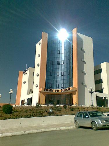
University SÃĐtif 1 FERHAT ABBAS Faculty of Sciences
DÃĐtail de l'auteur
Auteur abdallahi Ahmedou cheikh |
Documents disponibles écrits par cet auteur


 Ajouter le rÃĐsultat dans votre panier Affiner la recherche
Titre : Quality control in medical imaging Type de document : document ÃĐlectronique Auteurs : abdallahi Ahmedou cheikh, Auteur ; Saad Khoudri, Directeur de thÃĻse Editeur : Setif:UFA AnnÃĐe de publication : 2024 Importance : 1 vol (66 f.) Format : 29 cm Langues : Anglais (eng) CatÃĐgories : ThÃĻses & MÃĐmoires:Physique Mots-clÃĐs : Physique Index. dÃĐcimale : 530 - Physique RÃĐsumÃĐ :
The objective of our study is to compare the recommended tolerances with the test results
obtained from three types of equipment: radiology, mammography, and CT scanner. We aim
to assess the compliance of each device with current quality and safety standards by examining
the performances relative to the acceptable thresholds. This comparison will enable us to
identify any discrepancies and implement the necessary improvements to optimize the
reliability and safety of diagnostics.Note de contenu : Sommaire
Introduction GÃĐnÃĐrale : ....................................................................................................................... 1
Chapitre I : Appareils d'Imagerie : Radiologie, Mammographie, Scanner.
I.1 Introduction à lâimagerie mÃĐdicale................................................................................................ 2
I.1.1. Principe physique de radiologie ........................................................................................ 3
I.1.1.1 La production des rayons X.................................................................................................. 3
I.1.1.2 Tube à rayons X ................................................................................................................... 3
I.1.1.3 Cathode ............................................................................................................................... 4
I.1.1.4 Anode ................................................................................................................................. 4
I.1.2. Grille anti diffusant ........................................................................................................... 5
I.1.2.1 principe de fonctionnement ................................................................................................ 5
I.1.2.2 Facteur de bucky ................................................................................................................. 6
I.1.2. Principe physique de mammographie .............................................................................. 6
I.1.2.1 GÃĐnÃĐrateur à haute tension ................................................................................................ 6
I.1.2.2 La compression .................................................................................................................... 6
I.1.2.3 Lâexposeur automatique ...................................................................................................... 7
I.1.3. Principe physique de scanner ........................................................................................... 8
I.1.3.1 DÃĐtecteur............................................................................................................................ 8
I.1.3.2 Gantry (Cercle) :................................................................................................................... 8
I.2. Les modalitÃĐs en imagerie mÃĐdical ............................................................................................... 8
I.2.1 Radiographie ...................................................................................................................... 8
I.2.2 mammographie .................................................................................................................. 9
I.2.3 scanner ............................................................................................................................. 10
Fonction de transfert de modulation (FTM) .................................................................................... 11
Chapitre II : Protocol de control qualitÃĐ en imagerie mÃĐdical
â Ą.1. Introduction ............................................................................................................................... 12
â Ą.2. Les testes ....................................................................................................................................
â Ą.2.1 Radiologie (21) ................................................................................................................. 13
â Ą.2.2 mammographie (22) .......................................................................................................... 14
â Ą.2.3 Scanner (23) ...................................................................................................................... 15
Chapitre III : RÃĐsultats et discussion
Introduction .......................................................................................................................................
MatÃĐriel utilisÃĐ ...................................................................................................................................
Appareil radiographie....................................................................................................................... 18
â Ē-1 Radiographie marque comed : ......................................................................................... 18
A/ Clinique Dr aloune youssef ........................................................................................................... 22
1) Test dâalignement du champ lumineux avec le champ RX ................................................. 22
Methodes ................................................................................................................................. 22
2)Test rÃĐsolution bas et haut contraste ..................................................................................... 23
3) Etude de rÃĐpÃĐtabilitÃĐ de gÃĐnÃĐrateur ...................................................................................... 25
4) SensitomÃĐtrie ........................................................................................................................ 25
B/Clinique EPSP ............................................................................................................................... 26
1) VÃĐrification de la tension kVp ............................................................................................. 26
2)VÃĐrification du temps d'exposition ....................................................................................... 27
3)Test de facteur de bucky ....................................................................................................... 28
â Ē-2 mammographie .......................................................................................................................... 29
MÃĐthodes ................................................................................................................................. 33
2) Test de dose glandulaire moyenne ....................................................................................... 35
3) Test de rÃĐsolution spatial ...................................................................................................... 36
4) Utilisation de fantÃīme PE Hamed pour vÃĐrifier la qualitÃĐ dâimage ..................................... 38
â Ē-3 Scanner ......................................................................................................................................
â Ē-3-1QC scanner siemens ....................................................................................................... 39
â Ē-3-2 QC scanner Eclos Hitachi ............................................................................................. 45
â Ē-3-3 QC scanner Philips........................................................................................................ 53Côte titre : MAPH/0629 Quality control in medical imaging [document ÃĐlectronique] / abdallahi Ahmedou cheikh, Auteur ; Saad Khoudri, Directeur de thÃĻse . - [S.l.]Â : Setif:UFA, 2024 . - 1 vol (66 f.) ; 29 cm.
Langues : Anglais (eng)
CatÃĐgories : ThÃĻses & MÃĐmoires:Physique Mots-clÃĐs : Physique Index. dÃĐcimale : 530 - Physique RÃĐsumÃĐ :
The objective of our study is to compare the recommended tolerances with the test results
obtained from three types of equipment: radiology, mammography, and CT scanner. We aim
to assess the compliance of each device with current quality and safety standards by examining
the performances relative to the acceptable thresholds. This comparison will enable us to
identify any discrepancies and implement the necessary improvements to optimize the
reliability and safety of diagnostics.Note de contenu : Sommaire
Introduction GÃĐnÃĐrale : ....................................................................................................................... 1
Chapitre I : Appareils d'Imagerie : Radiologie, Mammographie, Scanner.
I.1 Introduction à lâimagerie mÃĐdicale................................................................................................ 2
I.1.1. Principe physique de radiologie ........................................................................................ 3
I.1.1.1 La production des rayons X.................................................................................................. 3
I.1.1.2 Tube à rayons X ................................................................................................................... 3
I.1.1.3 Cathode ............................................................................................................................... 4
I.1.1.4 Anode ................................................................................................................................. 4
I.1.2. Grille anti diffusant ........................................................................................................... 5
I.1.2.1 principe de fonctionnement ................................................................................................ 5
I.1.2.2 Facteur de bucky ................................................................................................................. 6
I.1.2. Principe physique de mammographie .............................................................................. 6
I.1.2.1 GÃĐnÃĐrateur à haute tension ................................................................................................ 6
I.1.2.2 La compression .................................................................................................................... 6
I.1.2.3 Lâexposeur automatique ...................................................................................................... 7
I.1.3. Principe physique de scanner ........................................................................................... 8
I.1.3.1 DÃĐtecteur............................................................................................................................ 8
I.1.3.2 Gantry (Cercle) :................................................................................................................... 8
I.2. Les modalitÃĐs en imagerie mÃĐdical ............................................................................................... 8
I.2.1 Radiographie ...................................................................................................................... 8
I.2.2 mammographie .................................................................................................................. 9
I.2.3 scanner ............................................................................................................................. 10
Fonction de transfert de modulation (FTM) .................................................................................... 11
Chapitre II : Protocol de control qualitÃĐ en imagerie mÃĐdical
â Ą.1. Introduction ............................................................................................................................... 12
â Ą.2. Les testes ....................................................................................................................................
â Ą.2.1 Radiologie (21) ................................................................................................................. 13
â Ą.2.2 mammographie (22) .......................................................................................................... 14
â Ą.2.3 Scanner (23) ...................................................................................................................... 15
Chapitre III : RÃĐsultats et discussion
Introduction .......................................................................................................................................
MatÃĐriel utilisÃĐ ...................................................................................................................................
Appareil radiographie....................................................................................................................... 18
â Ē-1 Radiographie marque comed : ......................................................................................... 18
A/ Clinique Dr aloune youssef ........................................................................................................... 22
1) Test dâalignement du champ lumineux avec le champ RX ................................................. 22
Methodes ................................................................................................................................. 22
2)Test rÃĐsolution bas et haut contraste ..................................................................................... 23
3) Etude de rÃĐpÃĐtabilitÃĐ de gÃĐnÃĐrateur ...................................................................................... 25
4) SensitomÃĐtrie ........................................................................................................................ 25
B/Clinique EPSP ............................................................................................................................... 26
1) VÃĐrification de la tension kVp ............................................................................................. 26
2)VÃĐrification du temps d'exposition ....................................................................................... 27
3)Test de facteur de bucky ....................................................................................................... 28
â Ē-2 mammographie .......................................................................................................................... 29
MÃĐthodes ................................................................................................................................. 33
2) Test de dose glandulaire moyenne ....................................................................................... 35
3) Test de rÃĐsolution spatial ...................................................................................................... 36
4) Utilisation de fantÃīme PE Hamed pour vÃĐrifier la qualitÃĐ dâimage ..................................... 38
â Ē-3 Scanner ......................................................................................................................................
â Ē-3-1QC scanner siemens ....................................................................................................... 39
â Ē-3-2 QC scanner Eclos Hitachi ............................................................................................. 45
â Ē-3-3 QC scanner Philips........................................................................................................ 53Côte titre : MAPH/0629 Exemplaires (1)
Code-barres Cote Support Localisation Section DisponibilitÃĐ MAPH/0629 MAPH/0629 Mémoire Bibliothèque des sciences Anglais Disponible
Disponible

